The Imaging & Optics Facility provides a full Image Acquisition Service for Slide-Scanning. One main advantages for using this service is that the acquisition is performed by Imaging and optics Facility staff – which can save time for your other research tasks. The service offers:
- Option to choose and combine different imaging modality (brightfield or fluorescence)
- Generation of overview scans for fast screening and sample navigation (see point 6)
- 2D or 3D imaging capabilities.
- Include high-resolution imaging of selected Regions Of Interest (ROIs), including shading correction & stitching
- Optional: scene splitting, further post processing and image analysis routine: deconvolution, segmentation
- Scanned images can be used for navigation on LSM confocal microscopes via ZEN Connect
The Slide Scanner Service makes use of the Zeiss Axioscan Z1. All specification in the form of available imaging modalities (brightfield, color and fluorescent), fluorecent excitation and objectives can be found at the system specification


Service Information
Ideal for larger batches: Histology or Fluorescent, samples.

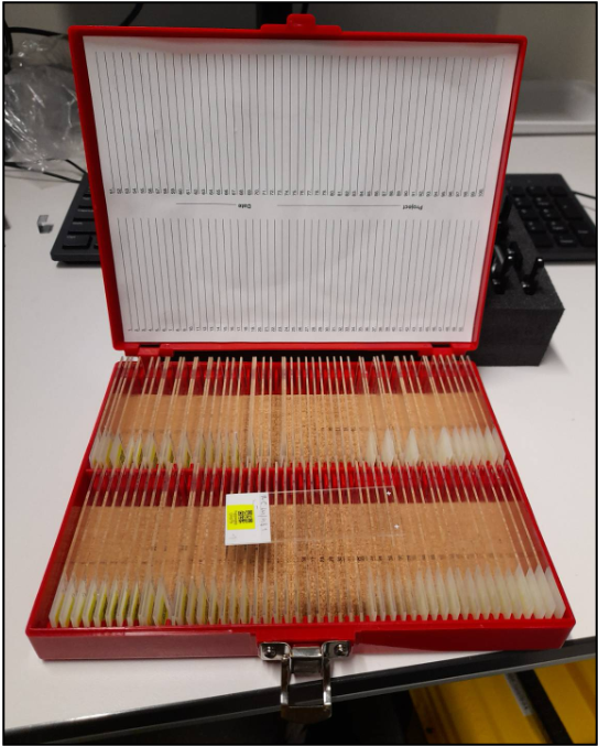
QR codes are provided as labels
 Multiple serial Sections can be placed on one slide
The sections are detected, or custom Regions Of Interest (ROI) can be defined to acquired multi-tile stack images

Z-projection of an 3-channel multi-tiled image stack