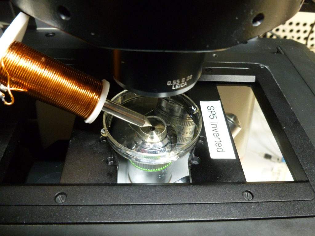
We have a magnetic tweezer setup that can be used on our Leica SP5 inverted confocal
The tweezer needs to be booked seperately in PPMS
For an introduction to the device please contact Doreen Milius
Magnetic Tweezers can be used for studying mechanical properties of biomolecules, tissues or cells
Magnetic tweezers are instruments that exert forces on magnetic particles (beads) through a magnetic field gradient.
Most commonly magnetic tweezers are used to study mechanical properties of biological macromolecules like DNA or proteins. Other applications are the study of the viscosity of intracellular fluids and of force-regulated processes in living cells. Forces are typically on the order of pico- to nanonewtons. Due to their simple architecture, magnetic tweezers are a popular biophysical tool.
The magnetic tweezer consists of a coil with a sharpened tip, which is made from an iron-nickel-alloy with a copper wire wrapped around it. The coil is connected to a power supply and controller which can be operated via a simple Matlab based program, where the strength of the magnetic field can be altered by applying different electric currents.
In the experiments, magnetic microparticles (beads) are injected into cells or whole organisms e.g. embryos. The tunable magnetic field coming from the coil is then used to manipulate magnetic beads whose position is measured with the help of time lapse microscopy.
Drosophila embryo injected with 4 µm beads
Zebrafish embryo injected with 2 µm beads