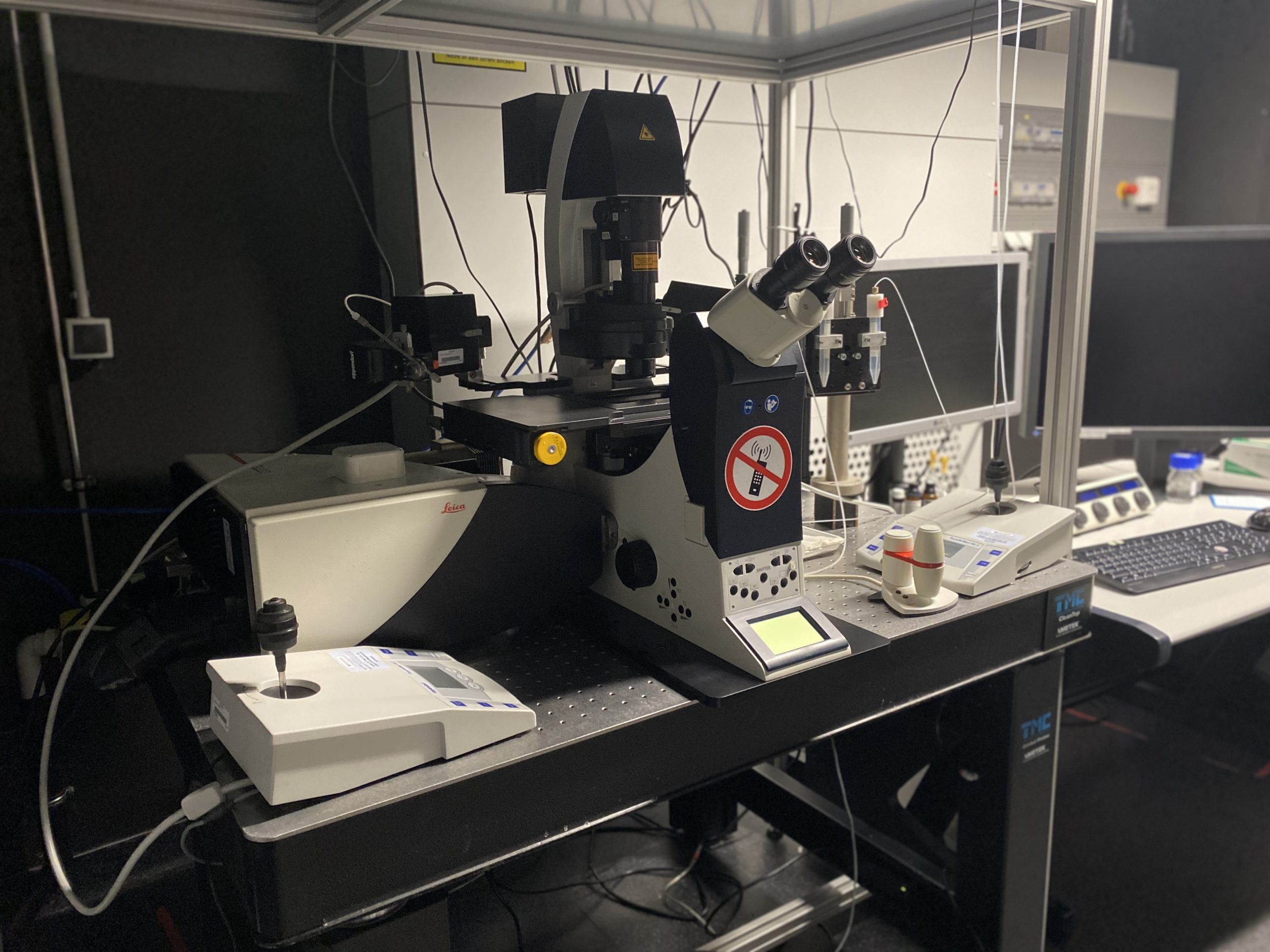
System has been replaced – please move ongoing experiments to new alternatives

Location
room: i04.U1.019:
Imaging modalities/features:
- Confocal
- Free emissions spectra selection
- Emission λ wavelength scans
- FRET and FRAP software
- Tandem scanner for ultra-fast imaging
- Micro-pipette aspiration
- Magnetic tweezers (book in PPMS)
Software: LAS AF 2.5.7.23225
Specification:
- DMi reseach microscope
- TCS scan unit
- Motorised Scanning stage
- Super Z Galvo Stage Type H
- Heated stage and objective heater (Pecon)
Light sources:
- p100 white, LED Light source (Cooled)
- Argon Laser lines 458, 488, 514,
- DMOD 561 nm
- DMOD 633 nm
Filters
- DAPI: Excitation: BP 350/50 Emission: BP 460/50
- FITC: Excitation: BP 480/40 Emission: BP 527/30
- RHOD: Excitation: BP 546/10 Emission: BP 585/40
Objectives (M25 thread):
- HC PL APO 20x/0.7 CS (11506513), WD=0.59 mm, D=0.17 mm
- HCX PL APO 40x/1.25-0.75 OIL CS (11506251), WD=0.1 mm, D=0.17 mm
- HCX PL APO 63x/1.4-0.6 OIL CS (11506188), WD=0.1 mm, D=0.17 mm
- HCX PL APO 63x/1.1 W CORR CS (11506279), WD=0.22 mm, D=0.14-0.18 mm)
- HC PLAN APO 10x/0.4 0.17/A2.2 (11506165), WD=2.2 mm, D=0.17 mm
- HC PL IRAPO 40x/1.10 W CORR, WD=0.63, D=0.14-0.18 mm
Detection Filters:
- NF 445
- NF 488
- NF 514
- NF 458/514
- NF 488/561/633
- NF 594
- NF 445/594
Detectors:
- 3x PMTs (free selection of detection range)
- 2x HyD SMD (free selection of detection range)
- 1 x Transmitted light detector (TLD)
Contact: Bioimaging Facility
Manual
Detailed description and manual: Leica_SP5_Inverted_confocal_microscope manual.pdf
