The Lattice Light Sheet Microscope (LLSM) is designed for long-term volumetric imaging at sub-cellular resolution. The system allows standard sample carriers, and is equipped with temperature, humidity and CO2 control. The Lattice Lightsheet technologies reduces phototoxicity and photobleatching, allowing monitoring of cellular processes for extended period (hours – to – days).
Description
su_spoiler title=”when to choose the ‘Lattice Lightsheet’ over the ‘Gaussian Lightsheet’ ” open=”no” style=”fancy” icon=”chevron” anchor=”” anchor_in_url=”no” class=””]
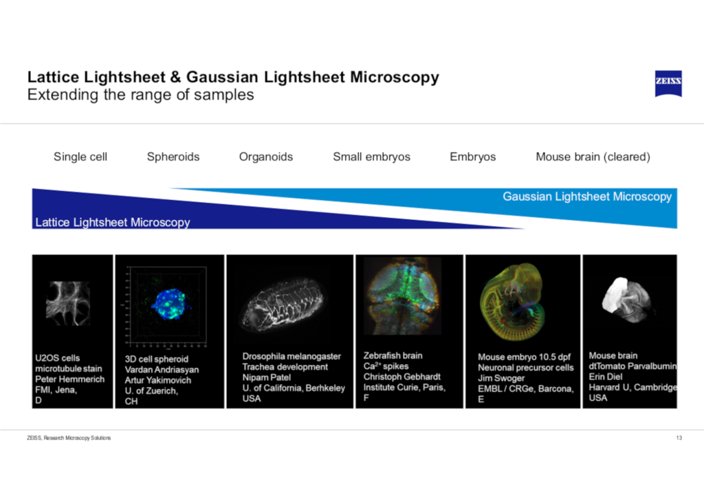
The Lattice Light Sheet Microscope takes over where there solution of the Gaussian light sheet (see our Zeiss Lightsheet 7) comes to its limits for sub-cellular imaging. The figure below gives an impression of the feasible size ranges: your samples should not exceed the range limit of maximum 300 µm in XY and 200 µm in Z. Samples can be mounted on well plates, MatTek dishes and slides (cover glass no. 1.5).
Advantages of LLSM
- Very fast
- Gentle illumination of the cells and fluorophores
- Gives enhanced resolution in the confocal range
- Inverse configuration allows the use of standard sample carriers
- Sample alignment and leveling is performed automatically by the system
- Automated immersion media supply (water)
 The Zeiss Lightsheet 7 is fully equipped for larger sample imaging, that include organoids, model organisms, cleared and expanded tissues.
Specification
- Multi-position & Tiling
- Versatile chambers and sample holders, with temperature, humidity and CO2 control.
- Additional piezo driven z-axis for fast imaging
- Transmitted Light (+ enhanced oblique contrast)
- Dual camera (2 x Hamamatus Orca Fusion sCMOS)
- Lattice Light sheets (typical): 500nm x 12µm ;650nm x 15µm; 750nm x 30µm; 1000nm x 30µm;1400nm x 85µm; 2000nm x 100µm
- Lateral resolution: 330nm x 330nm (+ Deconvolution: 290nm x 290nm)
- Axial resolution(depending on lightsheet): ranging from 500nm to 1000nm (+ Deconvolution: ranging
from 450nm to 900nm)
Objectives
The system is equipped with dedicated optics, and an automated water-immersion dispenser
- Excitation objective: 10x / 0.4, Water Immersion
- Meniscus lens
- Detection objective: 48x / 1.0, Water Immersion + motorized coverslip adjustment
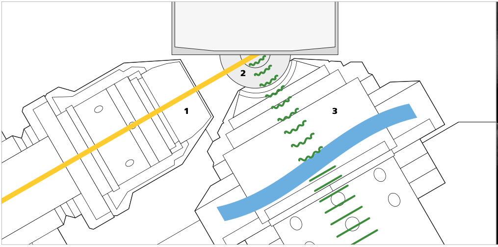
Schematic below shows the sample carrier and core optics module with excitation objective (1), meniscus lens (2) and detection objective with free-form optics (3).

Sample mounting
The system is optimized for conventional sample preparation techniques, and is equipped with temperature, CO2, O2, and humidity control. Please take note to use the correct cover slips (#1.5) !
- holder #1: petri dish 35-40mm
- holder #2: chamber slides 25 x 57mm
- holder #2: multiwell plates 85 x 127mm
Laser lines
The Lattice Light Sheet 7 is configured with the “high laser power laser module” to allow more flexibility and explore SMLM applications
- 488nm (14mW)
- 561nm (9mW)
- 638nm (13mW)
Dichroic / Camera Splitter:
- LP 565
- LP 640
Camera Filters:
| position |
Camera 1: direct light path | Camera 2: reflected light path |
| 1 | BP 570-620 + LP655 | BP 570-610 IR+ |
| 2 | BP 495-550 + LP655 | empty |
| 3 | BP 495-550 / BP 570-620 | BP 495-550 / BP 570-620 |
| 4 | LBF 405/488/561/642 | BP 500-550 IR+ |
| 5 | 1% ND | |
| 6 | empty | |
| 7 | BP 495-590 | |
| 8 | LP 570 |
Camera’s
- 2x Hamamatsu ORCA-Fusion sCMOS (> 82% QE und 6.5 µm pixel size)
Resources:
Additional information
| Building | i06 |
|---|---|
| Camera / Detector | sCMOS |
| Fluorescent Lightsource | Laser: 488±5 nm, Laser: 561±5 nm, Laser: 640±5 nm |
| System_Specification | Illumination: Fluorescence, Incubation: heating chamber, Incubation: heating stage, Incubation: CO2/N2-gas mixer (hypoxia), Automated Stage, Z-piezo / galvo Stage, Focus Stabilization, Dual Camera (see detectors), Automated Water Immersion Dispenser |
| Technology / Application | Time-lapse recording, In-vivo imaging |