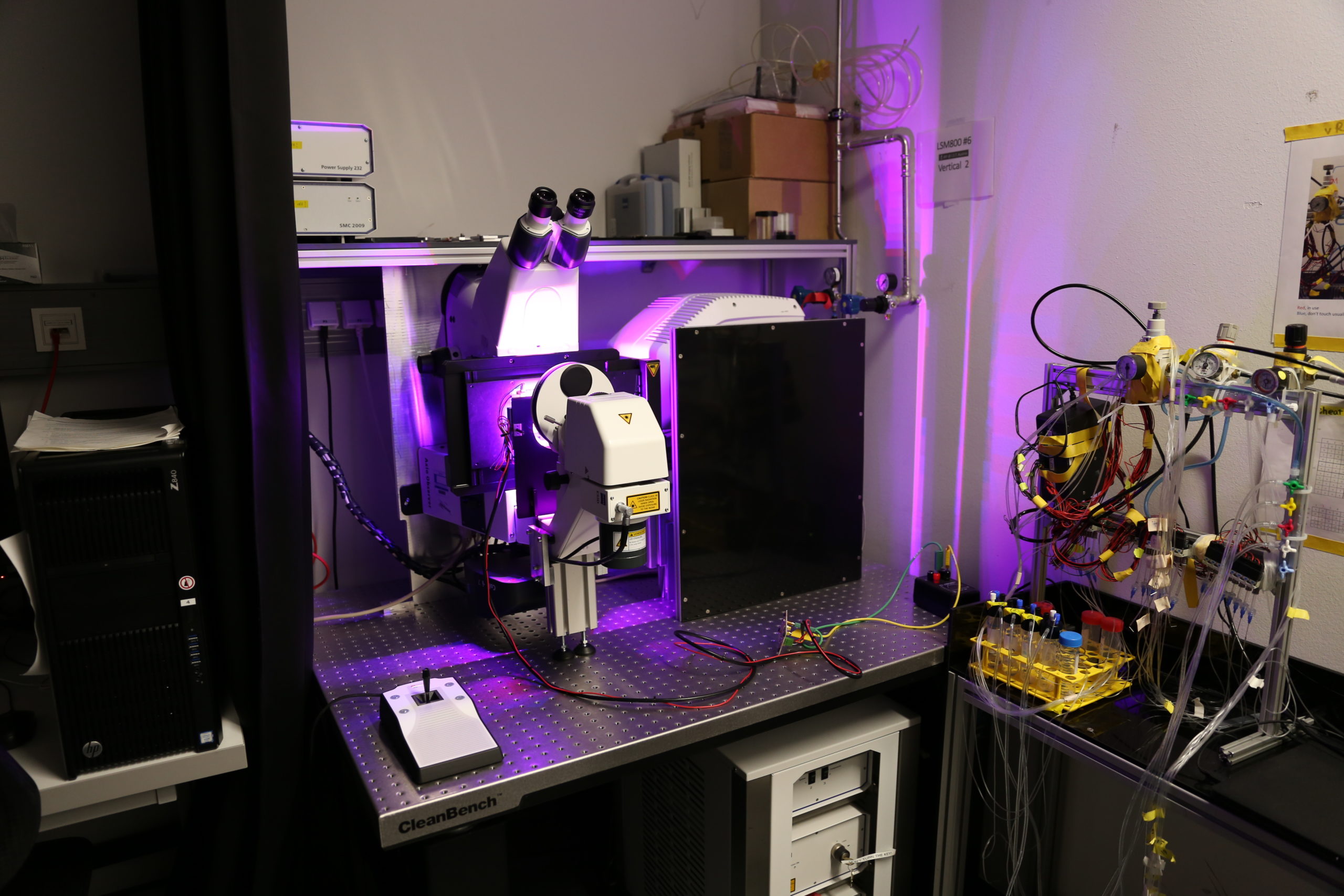
The vertical point scanning confocal microscope and root-tracker is a unique device that allows to image and track multiple samples / plant roots over a prolonged time in an upright position. This specific system provides a platform for both widefield and confocal imaging modalities.
Description
Location: room I04.O3.014 (BFB)
System Specification: Vertical confocal microscope
- 2x MA-PMT detectors: 2 channels simultaneously, more sequentially
- 1x GaAsP detector in confocal mode
- 1x CMOS camera (Axioscam 707 mono)
- Multiple position imaging
- Gravitation manipulation (rotation stage)
- (Root-)tracker
Light sources:
- Excitation Laser lines 405, 488, 561, 640 nm
- Fluorescent lamp: Zeiss Viluma 7-lines LED: 385/30; 423/44; 467/38; 508/20; 555/30; 591/27; 631/33; 735/40 nm
- ESID LED
Detectors:
- 2x MA-PMT: Multi Alkali Photomultiplier Tube detectors with free choice of the spectral range. (Uses a tunable short-pass and band-pass filter in front of the PMTs
- 1x GaAsP PMTs: Gallium Arsenide Phosphide photo-multiplier tube → do not overexpose / saturate!
- 1x ESID+LED light source: Electronically switchable illumination and detection module
- 1x cMOS camera: to be used for widefield-mode, in combination with the Viluma light source
Motorized stage for multiple position imaging → only inserts that belong to the system are allowed!
- Motorized stage for multiple position imaging
- Multi-slide holder, dish holder, single slide holder
Objectives (M27 thread)
- Plan-Apochromat 10x/0.45 , WD=2.1 mm – (420640-9900-000)
- Plan APOCHROMAT 20x/0.8 , WD=0.55 mm (D=0.17 mm) – (420650-9902-000)
- C-Apochromat 63x/1.2 Water, WD=0.28mm (CG=0.17mm) (421787-9970-799)
Software: Zen 3.12
System manual and additional material is available on SeaFile (login required).
- FIJI script to convert the image-sequence from tracker to concatenated files per position can be found here
- More details about the system can be found here: https://elifesciences.org/articles/26792
Additional information
| Building | i04 |
|---|---|
| Camera / Detector | MA-PMT, ESID, sCMOS |
| Fluorescent Lightsource | Laser: 405±5 nm, Laser: 445±5nm, Laser: 488±5 nm, Laser: 561±5 nm, Laser: 640±5 nm, LED: 355-365nm, LED: 430-450nm, LED: 545-555nm, LED: 640-650nm |
| System_Specification | Illumination: Fluorescence, Illumination: Bightfield, Automated Stage, Microfluidics |
| Technology / Application | FRAP, FRET, Time-lapse recording, Multi-position, Sample Stabilization / (Root-)Tracker, In-vivo imaging, Photoactivation / -conversion, Super Resolution (SR), SR: Airyscan (140nm) |
| Custom Tools / Inserts | Root-Chip Fluidics stage-Insert compatible (Lab) |


