The bioimaging facility at I.S.T. Austria is hosting a demonstration of the ZEISS Lattice Lightsheet 7 system from March 29th until April 2nd, 2021. The setup will be available in room i21.U1.027 (Basement Lab Building West).
Edit: Zen 3.3 LLS7 software has been installed on Workstation 1. You can use the software to deskew the raw data or deconvolve the Z-stacks. We also suggest that you look at the data in your favorite image analysis program.
A (remote) seminar is provided by Dr Jacques Paysan (Product & Application Specialist) on Monday, March 29, 2021. The seminar includes a brief explanation of the differences between Gaussian Lightsheet & Lattice Lightsheet imaging applications.[link to the seminar]
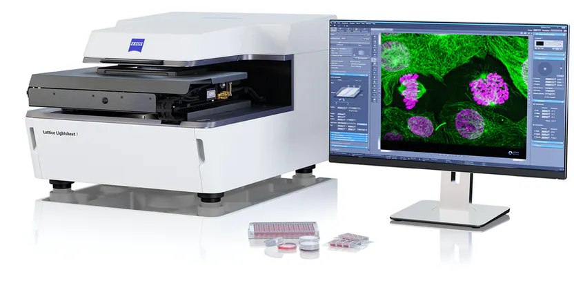
ZEISS Lattice Lightsheet 7 makes light sheet fluorescence microscopy available for live-cell imaging at subcellular resolution – while also allowing you to use your standard sample carriers. With this automated, easy-to-use system, volumetric imaging of subcellular structures and dynamics over hours and days with the best protection from photodamage becomes available to everyone. Discover the dynamics of life in the unprecedented depth of detail – with the ease you never imagined possible!
Info: ZEISS Homepage – Lattice Lightsheet 7
What are the advantages of using this system?
- Very fast
- Gentle illumination of the cells and fluorophores
- Gives enhanced resolution in the confocal range
- Inverse configuration allows the use of standard sample carriers
- Sample alignment and levelling is performed automatically by the system
- automated immersion media supply (water)
What kind of sample can I bring?
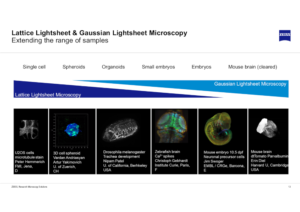
With this system, you can image live samples over long periods which makes it interesting for all kinds of over-night experiments with sensitive cells. The sample size can range from adherent cells to small organoids, zebrafish embryos and oocytes. Imaging of developing plants and plant seeds is also feasible.
Please note that the LLS7 cannot image beyond that size. The figure below gives an impression of the feasible size range. Your samples should not exceed the range limit of maximum 300 µm in XY and 200 µm in Z. Samples can be mounted on well plates, Mattek dishes and slides (cover glass no. 1.5).
Although the system comes with a gas mixer we do not have access to a CO2 tap in the demo room. Please supplement the medium accordingly.
WORKSHOP PROGRAM:
Seminar: Monday, March 29th 1pm→ The seminar include a brief explanation about the difference between Gaussian Lightsheet & Lattice Lightsheet imaging applications and requirements![link to the seminar]
Demo test sessions:
We offer morning and afternoon slots between March 29rd and April 2nd*. Please see the following schedule:
| Mon 29th | Tue 30th | Wed 31th | Thu 01st | Fri 02nd | |
| 9:30h-11:30h | BIF | David BABIC
(Friml) |
– | MedUni
(External) |
Mojtaba Tavakoli (Danzl) (AnHa) |
| 11.30h | lunch | lunch | lunch | lunch | Stephen Barratt
(De Bono) (AnHa) |
| 12:30-15h | 13:00 seminar by Jacques Paysan | Gayathri Singaraju
(Heisenberg) |
Xin Tong (Heisenberg) (AnHa) |
Ingrid DE VRIES
(Sixt) |
BIF |
| 15h-17h | Caterina Giannini | Suyash Naik
(Heisenberg) |
Saren Tasciyan
(Sixt) (AnHa) |
Nikhil Mishra
(Heisenberg) |
* We are awaiting feedback from the company for the defined times for the demo-slots
Requests for demo slots:
Please provide the following information to bioimaging@ist.ac.at:
- Preferred time slot
- Short description of your sample: fluorophores
- Sample carrier
- Temperature (?)
Specifications and components:
Objectives:
Illumination 13.3x /NA 0.4
Detection 44.83x /NA 1.0
Illumination
LED (white and red) for transmitted light
Laser (488 nm, 561 nm, 640 nm) for reflected light and epi-fluorescence
Camera
Pco.edge 4.2 CLHS
Filters
LBF 405/488/561/642
BP 495-550 /BP 570-620
BP 495-550+LP655
BP 570-620+LP655
EF LP 570
EF LP 488
Empty
ND filter
The ZEISS Implementation of Lattice Lightsheet Microscopy
During the development of Lattice Lightsheet 7, ZEISS gave special attention to user-friendliness and compatibility with conventional sample preparation techniques. An inverse configuration is the most important prerequisite to allow the use of standard sample carriers for high-resolution microscopy. The challenges resulting from an inverse configuration are mainly refractive index mismatches as fluorescence is emitted from the sample, passes through aqueous cell culture media, a tilted glass coverslip and water immersion, then into the detection objective.
Unrivalled ZEISS Optics

Resource: all information was taken from the supplier page